
 Provider
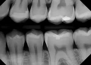
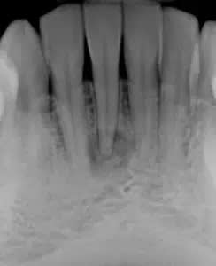
You may wonder why you orthodontist wants to take an updated x-ray when your dentist may have taken one recently. The x-rays that are taken most often by your dentist are intra-oral x-rays, taken inside the mouth used to focus on one or a few teeth to reveal the health of the enamel, inner canals, and roots. This allows your dentist to determine if there is decay or infection present. These x-rays are called bitewings (that show a sequence of teeth) and periapical (that primarily focus on one tooth).
Orthodontists are also dentists, and although they focus on different things, they share the responsibility of identifying any abnormalities that could be present such as abscesses, tumors, cysts and more. Although rare, identifying such pathologies is a very important step for patients to be referred for treatment.
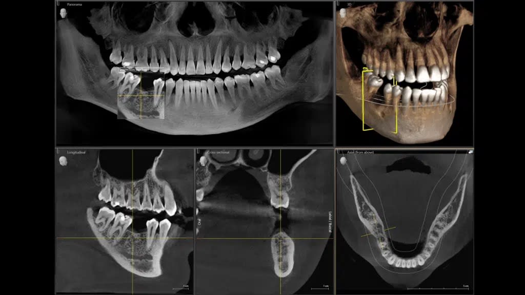
The type of x-rays that your orthodontist takes are extraoral x-rays, which are not as detailed as the bitewing and periapical x-rays that your dentist would take but are used to focus on the jaw and skull rather than specific teeth. These x-rays allow your orthodontist to monitor your overall jaw development, as well as see impacted, missing, extra and unerupted teeth. These images can be both two dimensional or three dimensional and can show soft tissue, nerves and bone. Being able to see jaw position, tooth position, and tooth root position is essential for your orthodontist to create your treatment plan.
Orthodontists are also dentists, and although they focus on different things, they share the responsibility of identifying any abnormalities that could be present such as abscesses, tumors, cysts and more. Although rare, identifying such pathologies is a very important step for patients to be referred for treatment.
The type of x-rays that your orthodontist takes are extraoral x-rays, which are not as detailed as the bitewing and periapical x-rays that your dentist would take but are used to focus on the jaw and skull rather than specific teeth. These x-rays allow your orthodontist to monitor your overall jaw development, as well as see impacted, missing, extra and unerupted teeth. These images can be both two dimensional or three dimensional and can show soft tissue, nerves and bone. Being able to see jaw position, tooth position, and tooth root position is essential for your orthodontist to create your treatment plan.
Book Your Complimentary Consultation
We would love to schedule your complimentary consultation, where our team will provide you with options to treat your condition and start your journey to a healthy, functional smile.